| McSwiggen & Associates |
 |
 |
| /Services |

McSwiggen & Associates offers a broad range of services to its customers. These include:• Analytical Services
• Petrographic Investigations and Optical Microscopy
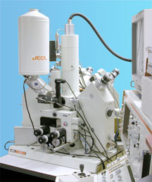
• Training and Support for JEOL Electron Microprobes
• Research on Geological or Electron Microprobe and Microscopy Topics
Quantitative Analysis Map Analysis Phase Analysis Line Analysis |
JEOL 8600 Electron Microprobe. |
Qualitative Analysis
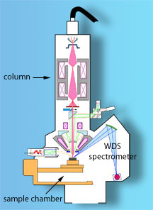
Qualitative analysis is used to determine what elements are present in specific regions of the sample. Qualitative analysis can be done either rapidly using an energy dispersive spectrometers, or with 10x the energy resolution using wavelength dispersive spectrometers (see Tech Notes: WDS vs EDS). Electron Imaging |
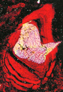
Polarized light microscopy is still one of the most fundamental ways to investigate the mineralogy of a rock. It still provides one of the fastest and least expensive methods for determining the minerals present and their textural relationships, and from this determine how the rock formed. Optical microscopy can also detect, in certain circumstances, very subtle compositional changes simply from observing changes in their optical properties. McSwiggen & Associates provide both transmitted and reflected light microscopic evaluations. |
|
||
Photomicrograph of a micronodule from |
 |
McSwiggen & Associates provides training and support to new JEOL electron microprobe owners. We cover the basic software operations and analytical procedures. We also run advanced training classes at the UCLA Microprobe Laboratory. These courses are customized to meet the needs of those registered, but typically cover advanced analytical procedures, methods for dealing with problem samples, instrument calibration techniques, and more in-depth exploration of the physical concepts involved in X-ray microanalysis. Links to other JEOL Electron Microprobe Labs
Schematic diagram of a JEOL |
|
| back to top |
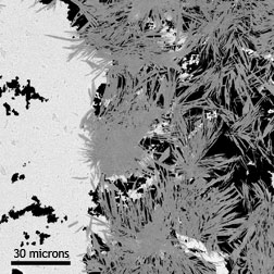
McSwiggen & Associates has been and continues to be involved in many different research projects related to the geology of Minnesota, and in the development of analytical procedures and methods, including:
Greenalite clast (white) in contact |
 |